Categories
North Gujarat's premier dedicated glaucoma super-specialty centre.

Computerized Automated Perimetry for mapping peripheral and central visual sensitivity.
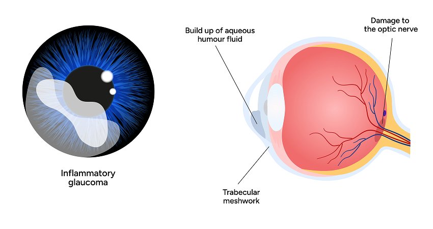
While OCT imaging reveals microscopic structural loss, Computerized Automated Perimetry (Visual Field Testing) is the definitive method for evaluating the functional consequence of glaucoma. Glaucoma typically attacks the peripheral visual field first, leaving central sharp vision untouched until late in the disease.
Atharva Glaucoma Centre is equipped with the gold-standard Humphrey Field Analyzer (HFA) utilizing the scientifically validated SITA (Swedish Interactive Thresholding Algorithm) strategy. Visual field testing maps each eye's differential light sensitivity, identifying subtle scotomas (blind spots) and guiding timely therapeutic adjustments.
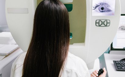
During the test, the patient sits comfortably in front of a calibrated illuminated bowl, with one eye occluded. The patient gazes at a central fixation light while micro-bursts of light with varying intensities appear at different coordinates across the field of vision.
Whenever a light stimulus is perceived, the patient clicks a handheld trigger. The computer cross-references responses against age-matched statistical baselines, generating greyscale deficit maps, pattern standard deviations (PSD), and Visual Field Index (VFI) percentages that quantify remaining visual function.
Our perimetry software features Guided Progression Analysis (GPA), which tracks multiple sequential visual field tests over months and years.
By filtering out temporary fluctuations caused by fatigue, GPA alerts Dr. Gandhi to true, statistically confirmed visual deterioration, allowing treatment escalation before significant vision is lost.
Why perimetry remains mandatory for every glaucoma patient:
Perimetry requires sustained focus because many lights are intentionally presented at the threshold of visibility—meaning you will miss about half the lights, which is completely normal.
You do not wear your own spectacles. Instead, our clinical team inserts a custom trial lens matched to your exact refractive correction into the perimeter's viewing aperture.
Newly diagnosed patients typically undergo 2 to 3 visual field tests in the first year to establish an accurate baseline, followed by annual or bi-annual monitoring depending on disease stability.