Categories
North Gujarat's premier dedicated glaucoma super-specialty centre.

High-resolution digital stereoscopic imaging of the optic nerve head and peripapillary retina.
The optic nerve head (optic disc) is where over one million retinal nerve fibers converge before traveling to the brain. In glaucoma, pathologically elevated intraocular pressure compresses these fibers, causing optic cup enlargement, neuroretinal rim thinning, and microvascular hemorrhages.
Digital Stereoscopic Disc Photography provides true-color, high-magnification photographic documentation of the optic nerve head and adjacent retinal architecture. Unlike clinical sketches or verbal notes, photographs provide an objective, permanent visual record that can be side-by-side compared decades into the future.
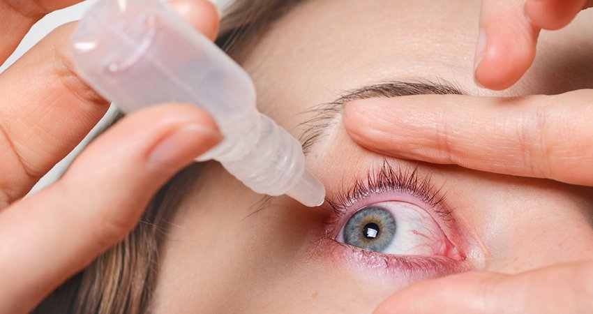
Following pupil dilation, a specialized high-resolution digital fundus camera captures stereoscopic image pairs of your optic discs under controlled illumination. The camera records subtle details such as cup-to-disc ratio, rim notch formation, baring of circumlinear vessels, and nerve fiber layer bundle defects.
Dr. Amola Gandhi evaluates these calibrated images alongside prior sessions. Subtle clinical markers—such as Drance disc hemorrhages, which indicate active glaucomatous damage—are readily identified, allowing proactive medical intervention before visual field loss occurs.
While OCT delivers mathematical thickness measurements, it cannot replace direct visualization of optic disc color, pallor, lamina cribrosa pores, and peripapillary atrophy zones.
Combining high-definition digital disc photography with automated perimetry and OCT offers a robust triple-check diagnostic standard for world-class glaucoma care.
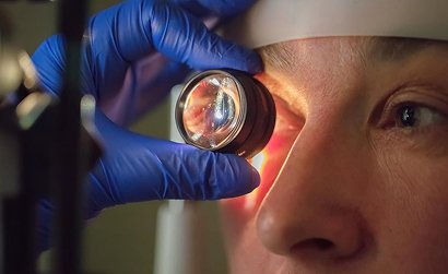
Why photographic documentation is vital for your lifelong ocular health:
Yes. Dilating the pupil ensures a wide optical aperture, allowing the camera to capture panoramic, high-definition photographs of the optic nerve without peripheral shadow distortion.
No. The camera flash uses a safe, momentary medical-grade xenon or LED burst that causes a brief after-image lasting only a few seconds without any harmful effect.
Absolutely. All digital images are archived in your electronic medical file at Atharva Glaucoma Centre and can be exported for medical records or referrals.