Categories
North Gujarat's premier dedicated glaucoma super-specialty centre.

Specialized gonioprism examination of the anterior chamber drainage angle to classify glaucoma types.
Due to the optical phenomenon of total internal reflection, the cornea prevents an examiner from viewing the eye's internal drainage pathway directly through an ordinary slit-lamp. Gonioscopy is the indispensable clinical technique that overcomes this barrier using a specialized gonioprism lens placed on the anesthetized eye.
Gonioscopy allows direct visual inspection of the anterior chamber angle—the crucial junction where the cornea and iris meet, containing the trabecular meshwork and Schlemm's canal. Performing gonioscopy is mandatory to definitively classify glaucoma as Open-Angle, Narrow-Angle, or Angle-Closure.
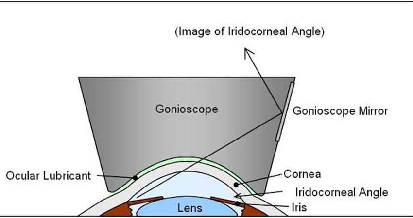
After applying topical numbing drops, Dr. Amola Gandhi gently rests a multi-mirror gonioprism (such as a Zeiss or Goldmann lens) on the cornea with a coupling fluid. Light beams are mirrored across the 360-degree circumference of the angle.
The examiner systematically grades all four anatomical landmarks: the Schwalbe line, trabecular meshwork, scleral spur, and ciliary body band. Furthermore, Dr. Gandhi performs dynamic indentation gonioscopy to differentiate between reversible appositional angle closure and permanent peripheral anterior synechiae (PAS).
In Asian populations, angle-closure glaucoma is particularly prevalent and significantly more aggressive than open-angle glaucoma. Undetected narrow angles can suddenly close, triggering excruciating ocular pain, nausea, and irreversible blindness within hours.
Gonioscopy identifies occludable angles in asymptomatic patients, allowing Dr. Gandhi to perform preventative Nd:YAG laser iridotomy before a blinding emergency ever occurs.
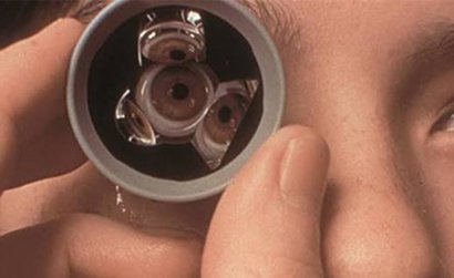
Critical clinical insights provided exclusively by gonioscopy:
Yes, the lens rests gently on the clear cornea. However, thanks to effective topical anesthetic eye drops, you will not feel pain—only a mild sensation of contact.
While AS-OCT provides helpful cross-sections, gonioscopy remains the clinical gold standard because it allows dynamic manipulation (indentation) and direct visualization of pigment, blood vessels, and tissue adhesions.
The numbing effect wears off within 15 to 20 minutes, and any mild blurriness caused by the lubricating gel clears after gently blinking or rinsing with artificial tears.