Categories
North Gujarat's premier dedicated glaucoma super-specialty centre.

Spectral-Domain Optical Coherence Tomography scanning the optic nerve and retinal nerve fiber layer.
Optical Coherence Tomography (OCT) has revolutionized modern ophthalmic diagnosis by functioning as an optical ultrasound that delivers micron-level, cross-sectional views of the living retina. In glaucoma, damage manifests first as the progressive thinning and loss of retinal ganglion cell axons in the Retinal Nerve Fiber Layer (RNFL).
At Atharva Glaucoma Centre, our advanced Spectral-Domain OCT platform captures thousands of A-scans per second without touching the eye. This provides quantitative, color-coded topographic thickness maps of the optic disc, peripapillary RNFL, and macular ganglion cell complex (GCC), detecting glaucoma years before visual field tests detect blind spots.

During the scan, low-coherence near-infrared light is directed into the pupil. The light reflections are captured with interferometry to construct high-resolution 3D anatomical models of the optic nerve head, cup-to-disc contours, and neuroretinal rim volume.
The acquired measurements are compared instantaneously against an extensive age-matched normative database. Areas with significant axonal thinning are flagged in yellow (borderline) or red (outside normal limits), giving Dr. Amola Gandhi objective data to detect pre-perimetric glaucoma.
Over 50% of the eye's retinal ganglion cells are concentrated within the central macula. Macular Ganglion Cell Complex (GCC) analysis scans the three inner retinal layers where early glaucomatous damage frequently begins.
This dual-scanning strategy (peripapillary RNFL plus macular GCC) provides unprecedented sensitivity, ensuring early diagnosis and accurate monitoring of therapeutic response over time.
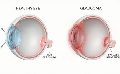
Why Spectral-Domain OCT is indispensable for modern glaucoma evaluation:
No. OCT uses harmless, low-energy near-infrared light waves similar to standard optical photography, making it 100% radiation-free and completely safe.
While modern high-speed OCT machines can scan through undilated pupils in many patients, mild dilation may be recommended if pupils are small or mild cataracts are present to obtain optimal scan clarity.
Stable glaucoma patients typically undergo OCT scans once every 6 to 12 months. Patients with suspected progression or borderline findings may be imaged more frequently.