Categories
North Gujarat's premier dedicated glaucoma super-specialty centre.

Multi-parameter clinical screening and structural evaluation for precise glaucoma diagnosis.
Glaucoma is frequently termed the "silent thief of sight" because it typically progresses painlessly without noticeable visual symptoms until irreversible optic nerve damage has already occurred. A Comprehensive Glaucoma Workup is an exhaustive, multi-step clinical assessment specifically engineered to detect microscopic structural changes and subtle functional vision loss at the earliest possible stages.
At Atharva Glaucoma Centre in Mehsana, Dr. Amola Gandhi utilizes state-of-the-art diagnostic instrumentation to deliver a meticulous baseline evaluation. This unified assessment combines precision intraocular pressure checks, corneal thickness adjustments, angle grading, stereoscopic nerve head examination, and automated visual field plotting, empowering us to formulate an individualised, sight-saving treatment strategy.
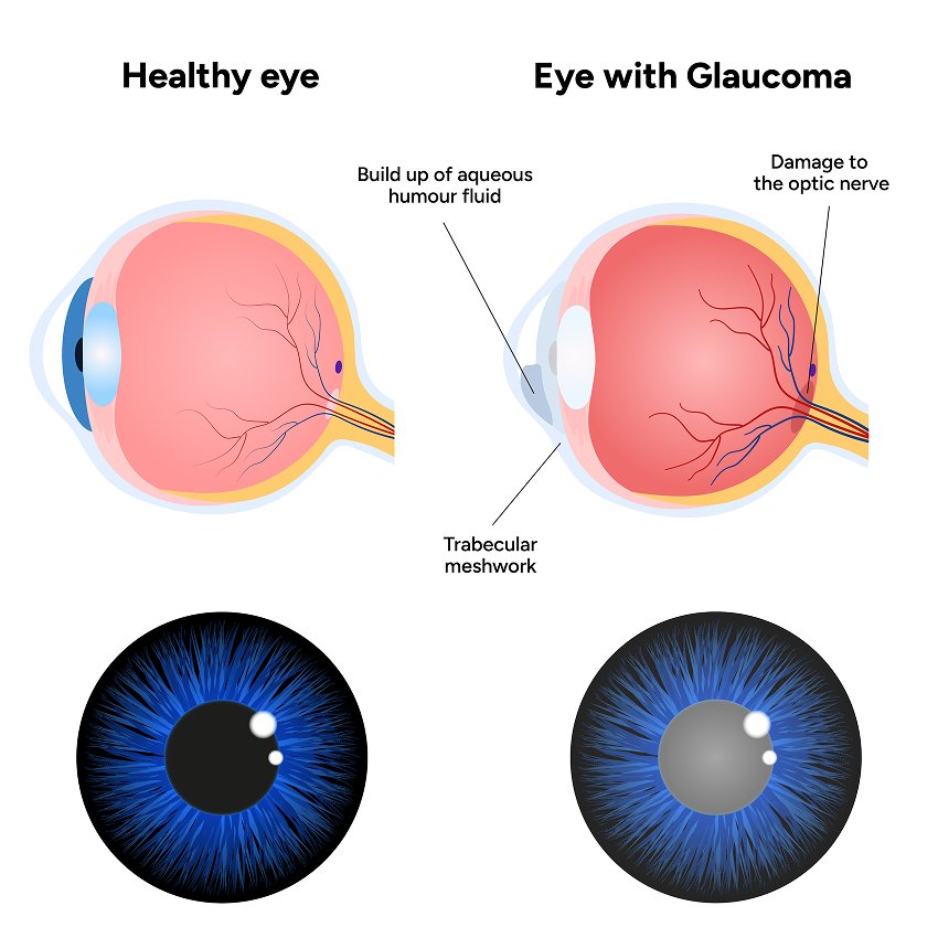
The workup begins with an in-depth medical history review and non-contact screening, followed by high-precision Goldmann applanation tonometry to establish your baseline intraocular pressure. We perform corneal pachymetry to assess corneal thickness, ensuring pressure readings are calculated with clinical accuracy.
Next, specialized gonioscopy is conducted with a four-mirror gonioprism to inspect the anterior chamber angle architecture and determine whether the drainage angle is open, narrow, or occluded. Following pupil dilation, stereoscopic optic disc photography and optical coherence tomography (OCT) capture high-resolution cross-sections of the retinal nerve fiber layer (RNFL), establishing a definitive reference for lifelong monitoring.
Modern glaucoma management demands reproducible, high-definition data. By integrating automated Humphrey visual field perimetry with spectral-domain OCT imaging, our centre pinpoints sub-clinical axonal loss years before symptoms appear.
This comprehensive baseline eliminates guesswork, distinguishes physiological large cups from early glaucomatous cupping, and ensures patients receive targeted therapies only when clinically warranted.
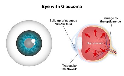
A complete evaluation provides vital diagnostic clarity that standard basic eye exams cannot match:
A full evaluation generally requires approximately 90 to 120 minutes. This allows sufficient time for pupil dilation, specialized corneal and optic nerve imaging, and thorough physician consultation.
No. Topical anesthetic eye drops are gently instilled prior to any direct contact tests such as tonometry or gonioscopy, ensuring a completely painless and comfortable experience.
Individuals over 40 years old, anyone with a family history of glaucoma, high myopia, diabetes, long-term steroid medication use, or those told they have elevated ocular pressure should be screened annually.